WORKSHOPS

Community and Industry workshops info

Community

Several sessions which address key topics from image processing to advanced imaging, prepared and presented by your peers

Industry @ room

Series of presentations on technical advances and applications directly from manufacturers, will occur in a dedicated room

Industry @ booth

Series of presentations on technical advances and applications directly from manufacturers, will occur directly at the manufacturer's booth

Community Workshops

Speakers:

» Catarina M. Seabra
» Mohamed Edfawy 

Session Description:

In this workshop you will learn how to apply microscopy techniques to answer your questions related to neuroscience research, both in mouse brain and human-derived brain organoids. Using peripheral injections of AAVs expressing a fluorophore under the control of a neuronal promoter, well individualized cells can be found across the entire mouse brain within 4-6 weeks post-injection. We have employed this technique to characterize neuronal arborization, spine morphology and provide proof of principle on its usability towards advanced methods to dissect neuronal network activity or gene function.

The recent breakthrough of the development of brain organoids has now allowed researchers to study the function of brain development and circuit formation in a more physiological context that has a 3D structure. From immunohistochemistry to in-vivo calcium imaging, microscopy techniques have been vital to dissect the intricate processes of brain organoid development and maturation.  

Speakers:

» Arrate Muñoz Barrutia
» Daniel Sage
» Hugo Botelho
» Joaquim Soriano Felipe


Session Description:

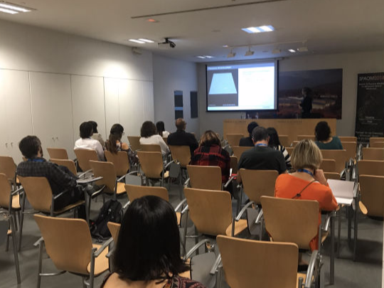
» Bridging Deep Learning to ImageJ - Arrate Muñoz Barrutia & Daniel Sage

Machine learning (ML), and in particular, Deep Neural Networks (DNN), have become an inflexion point in many areas of scientific research. In the case of biomedical image analysis, these new techniques provide significant improvements in most of the tasks such as denoising, super-resolution, segmentation, detection, tracking, response prediction or computer aided diagnosis.

Nonetheless, the use of DNN models requires previous programming knowledge and expertise, which makes them unapproachable to the general public. Therefore, the spread of this technology to the scientific community is strongly limited. In this workshop, we present a friendly interface for the use of these models that have been developed in collaboration between EPFL and UC3M. This ready-to-use ImageJ/FIJI plugin has the potential to make available many of the powerful algorithms for image processing that are continuously being developed and published, enhancing research.

 - more info here:

     https://deepimagej.github.io/deepimagej/

     https://www.biorxiv.org/content/10.1101/799270v1

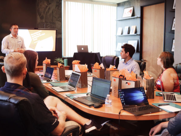
» Analysis of time lapse microscopy data with CellProfiler et al - Hugo Botelho

Automated microscopy is an invaluable tool in the research of complex cellular/organismal processes because it enables reproducible highly multi-dimensional imaging experiments (e.g. multi-position xyλt, where each position may represent a different experimental treatment across a multi-well plate). Scenarios like this are common in live cell assays or high content screening campaigns. Unfortunately, batch image analysis and proper result annotation (experimental metadata) may not be trivial for many researchers. Herein, I propose to demonstrate using CellProfiler in analyzing a small time lapse microscopy dataset with the goal of comparing the phenotypic response of intestinal organoids to gene editing and pharmacological intervention  https://github.com/hmbotelho/cellprofiler-practical2-NeuBIAS). The exercise will focus on how the following basic image analysis tasks are implemented in CellProfiler: background subtraction, object segmentation, feature extraction and results output). A pre-configured analysis pipeline will be provided for the users which wish to follow the demonstration in their own laptops. In the last few minutes I will show how the same
dataset could be analyzed in Knime or Fiji. Participants will not be able to follow along on their own computers (otherwise, the demonstration would be too lengthy) but pre-configured workflows/scripts will be made available as well.

Depending on available time and participant interest, advanced operations (e.g. object-level quality control, tracking and downstream data analysis) may also be demonstrated.

» ImageJ macro-based workflows produce better science


Science is hampered by reproducibility and economic issues. On one hand, lack of reproducibility is due to deliberate data biasing, lack of knowledge, inadequate supervising (usually from IP to Postdocs to students) and incomplete reports. On the other hand, economy limits data production and data sharing to the labs that can pay for: journal access and journal publication expenses, highly specialized tools (i.e. microscopes and image analysis software) and knowledge (i.e. courses and conferences). ImageJ macro-based workflows increase productivity and reproducibility, ease data sharing and are accessible to all users regardless of their technical background and economic status. Practical examples on the topic will be shown.
 

Speakers:

» Pedro Matos Pereira (@P_Matos_Pereira)

» Siân Culley (@SuperResoluSian)


Session Description:

Despite the widespread use of Super-Resolution techniques, most methods remain technically challenging and involve sophisticated hardware and/or software tools. In 2014, Expansion Microscopy (ExM) was introduced as an alternative to overcome this challenge. The concept is extremely simple: instead of super-resolving your target of interest you physically expand your sample in such a way that the structure is now above the diffraction limit (the expansion is 4 to 5 fold the original size) and image it on a conventional microscope. The original method served to deliver super-resolution information to researchers without access to a super-resolution system. However, ExM versatility was limited by technical details like the need for special labelling probes. Several improvements were made since then to enhance its applicability, such as introducing changes that made ExM compatible with conventional probes, like commercially available labelled antibodies and endogenous fluorescent proteins. Additionally, ExM can also be combined with classical super-resolution microscopy techniques effectively allowing to combine the resolution increase from both approaches. ExM has been applied to a variety of samples, from eukaryotic cells and tissues to pathogens such as bacteria or virus, making it an excellent alternative to classical super-resolution approaches.

Speakers:

» Sébastien Tosi
» Ignacio Arganda Carreras

Session Description:

Deep learning, the latest extension of machine learning, has pushed the accuracy of algorithms to unseen limits, especially for perceptual problems such as the ones tackled by computer vision and image analysis. This workshop will cover the foundations of the field, the communities organized around it, and some important tools and resources to get started with these techniques. Some successful applications of deep learning, especially in the field of bioimage analysis, will be presented and some hands-on will cover how to setup a working deep learning environment. No prior programming knowledge is required to follow the workshop.

Speakers:

» Julien Colombelli
» Gabriel Martins
» Jim Swoger
» Emilio Gualda


Session Description:

Mesoscopy is a new generic term that comprises the techniques allowing 3D fluorescence imaging of very large samples in toto (mm to centimeters). Mesoscopy has seen a fast pace of development recently due to improvements in light-sheet microscopy, optical tomography, and tissue clearing techniques. Because of the small offer of commercial systems to do mesoscopy, several labs have, in the past 5-6 years, begun developing DIY and even open source solutions for implementing these techniques, for example the openSPIM, OpenSpin microscopy, OPenT, MesoSPIM, LegoLISH or OptiSPIM.

In this small workshop we will demonstrate the principles of light sheet and optical tomography, some of the subsystems necessary to assemble a working mesoscope and a brief demo of how 3D mesoscopic datasets are acquired. 

Speakers:

» Erin Tranfield
» Mafalda Silva
» Ana Laura Sousa 

Session Description:

Correlative Light and Electron Microscopy: Introduction to experimental
procedures, as well as tricks and tips for successful experiment planning and execution.
 
Session Program: 

1. Intro to the different kinds of CLEM and some of the challenges of CLEM
2. CLEM applications: how to answer biological questions 
3. Discussion about how to start a project and when CLEM is an appropriate
4. Demonstration of CLEM tools

Speakers:


» Siân Culley (@SuperResoluSian)

» Pedro Matos Pereira (@P_Matos_Pereira)

Session Description:

In this session, we will look at Fiji workflows for single molecule localization microscopy (SMLM) data. The main points that we will cover are:

1. Analysis of sparse blinking datasets: the ‘classic’ SMLM dataset
• Use of QuickPALM and ThunderSTORM to localize individual molecules
• Interpretation of particle tables
• Drift correction
• Rendering and visualisation of images

2. 3D SMLM data sets
• Different types of 3D calibration data
• Localization and visualisation in ThunderSTORM

3. Dense blinking datasets: the ‘tricky’ SMLM dataset
• Use of HAWK for pre-processing dense data
• Options in ThunderSTORM for fitting dense data
• Using SRRF for live-cell data

4. Multicolour datasets
• Chromatic aberration correction using NanoJ

5. Assessing data quality
• Using SQUIRREL to measure the resolution of images and to assess the quality of images.

Homework: To prepare for this session, participants are asked to install Fiji (https://imagej.net/Fiji/Downloads). We will be using the following plugins:
• QuickPALM (pre-packaged into Fiji)
• ThunderSTORM (installation instructions at: https://github.com/zitmen/thunderstorm)
• HAWK (https://www.coxphysics.com/ - copy the version 1.1 .jar file into the plugins folder in your Fiji install)
• NanoJ-Core, NanoJ-SRRF, NanoJ-SQUIRREL (In Fiji, go to Help>Update…>Manage Update Sites and check the boxes for NanoJ-Core, NanoJ-SQUIRREL and NanoJ-SRRF. Close this window, then press ‘Apply changes’

We will also be using various test data sets (which we will provide or the participants can bring their own) 

Speakers:

» María Victoria Gomez

.Instituto de Investigación Sanitaria Gregorio Marañón, (IiSGM), Madrid, Spain
.Centro de Investigación Biomédica en Red de Salud Mental (CIBERSAM), Madrid, Spain
.Departamento de Bioingeniería e Ingeniería Aeroespacial, Universidad Carlos III de Madrid, Spain.


» Montserrat Coll Lladó (EMBL Barcelona) 

» Miguel Lopes (CNC)

Session Description:

3D IMAGING: OPTICAL TISSUE CLEARING


3D imaging tools are emerging in the field of optics, bioengineering and biomedicine. Opacity precludes light penetration into the tissues, making it difficult to obtain accurate 3D pictures of an organ without slicing it. Optical tissue clearing techniques enable light penetration into tissues and solve the scattering issue, allowing visualization of the whole tissue without the need to section it. Through tissue clearing, 3D imaging of whole organs is bringing light to basic and clinical research, including diagnostics.


Different methods and protocols for optical tissue clearing have been developed. In this workshop we will learn the advantages and disadvantages of each of the following clearing methods:
» General optics concepts: scattering and transparency.
» Protocols and methods: advantages and disadvantages
» Immunostaining of whole organs
» BABB method
» Whole organ clearing with the CUBIC protocol:

    - Advantages of the CUBIC method. 

    - Latest versions and optimizations of the CUBIC method 

» Clearing for confocal and SPIM microscopy image acquisition
» Biomedical applications: animal and human tissues.


Industry Workshops @ room

Speakers:

Dr. Sebastien Peter, Senior Product Specialist at Olympus Europe

Session Description:

Fast super resolution imaging: principles & applications


We are proud to invite you to this session that will cover the latest developments on confocal spinning disk microscopy (CSDM). With the high sensitivity and speed required for live cell and tissue imaging, the NEW SoRa Microscope from Olympus provides super resolution imaging at high speed real-time. Sora is a revolutionary tool can break diffraction limit up to 120nm easily.
Enjoy the last Olympus development and bring your own samples to check the amazing capabilities from SoRa microscope.  

Speakers:

Dr Gerry Sexton, Carl Zeiss Iberia SL, España
Dr Soren Prag, Carl Zeiss Microscopy, Jena, Germany 


Session Description:

Live-Cell Imaging with LSM980 Multiplexing and Celldiscoverer7 with LSM900
In recent years live-cell imaging has evolved into the new standard for all fields of life science research. This spans from live-cell imaging analysis of subcellular structure in super-resolution single cell microscopy into large three-dimensional structures like organoids, tissue sections or small whole organisms. These requirements for live-cell imaging are a major focus in the latest developments from ZEISS.
Experimental conditions that are as close as possible to the natural environment, sparse labeling and gentle imaging conditions are prerequisites to produce representative results in modern imaging tasks - especially in the context of experiments that last for hours or days. Additionally, the need for increased throughput and ease of use without compromising on image quality and information density is constantly growing.
In the recent years, ZEISS proudly launched several systems with major focus on live-cell imaging, namely the LSM980 with Airyscan2, Celldiscoverer7 with LSM900 and Airyscan2, the Elyra7 super-resolution system, and Lightsheet.Z1.
At this workshop we will present the LSM980 with Airyscan2 and the Celldiscoverer7 with LSM900. Our latest LSM980 with Airyscan2 is the ideal confocal microscope for 4D imaging. The entire beam path is optimized for simultaneous spectral detection of multiple weak labels with the highest light efficiency. Add the new Multiplex mode for Airyscan 2 to get more imaging options for your experiments. You can now choose the perfect setup to gently image larger fields of view with superresolution in shorter acquisition times than ever before.
ZEISS Celldiscoverer 7 with LSM 900 and Airyscan 2 is an automated microscope system, which fuses the advantages of widefield and confocal imaging and profits from a unique optical concept. Its sophisticated incubation allows for the execution of complex long-term imaging experiments even by non-microscopy experts. The unparalleled sensitivity of the camera as well as the LSM detectors guarantee for the most gentle image acquisition when working with sensitive biological samples. Airyscan 2 with its new Multiplex mode, enables the acquisition of large volumes with high temporal and optical resolution. 

Speakers:

» tba

Session Description:

Advances in Image Processing Automation and machine learning in Amira Software for Life Sciences
Powerful image processing tools are essential to the modern microscopist’s workflow. Image sizes are growing as microscopes advance in quality and power, and with that the need for workflow automation is growing to be more important than ever.

Thermo Scientific™ Amira™ Software rises to this challenge with the inclusion of recipes for image stack processing. Recipes are a mechanism by which tedious workflows of unlimited complexity are recorded, documented, and customized for application to further datasets.
These recipes can be applied as a batch to 2D images comprising a stack, or as a sequence of 3D image processing operations. Saving a recipe as a simple text file allows the quick and easy transfer of expertise between colleagues or research sites via email. Results of recipes can include processed images, segmentation results, statistical measures, meshed surfaces and volumes, and more.

During the workshop, the recipe creation process will be explained and demonstrated for segmentation of a life sciences dataset. Follow creation, the recipe will be applied on further datasets. We’ll describe in detail how automation of this segmentation process eliminates the main bottleneck of the image processing workflow and enables researchers to more quickly reach the results that push their research forward. In addition texture classification based on machine learning will be demonstrated. 

High-Speed Confocal Platform

Classical confocal imaging means a difficult choice between speed, sensitivity and resolution. Getting through Dragonfly specifications and examples, we can approach new confocal imaging to super resolution and giving opportunities to explore new microscopy solution.

Top tips for successful high-content screening assay with 3D models

Whether you’re familiar with high-content screening and are looking to exploit the increased physiological relevance of complex 3D cell models, or you want to take your analysis of 3D cell models to the next level, migrating from simple microscopy to a high-content approach, you’ll need the right tools and strategies to overcome the challenges these models present.
Challenges include:

• Difficulties of growing consistent and reproducible 3D cultures
• Generation of high-quality images from large, thick cell samples
• Long imaging times and management of the huge volumes of data from 3D cell screens
• Analyzing responses of complex models in three dimensions
• Having to transfer data between software packages to enable 3D analysis

In this seminar we will provide our top tips for running a successful high-content screen using a 3D cell model.

Industry Workshops @ booth

New 3D analysis and machine learning tools to optimize High Content Analysis

GE Healthcare launched new modules for IN Carta image analysis software.

VoluMetrics will provide a complete solution for 3D measurement and Phenoglyphs an innovative machine learning based solution for classification that will make you win a lot of time. Participating at this workshop will show you all the power of these new tools.

The benefits and optimisation of CoolLED light sources

LED-based fluorescence illumination systems for use in microscopy have been available for over 10 years. This workshop, presented by Jo Whetstone and Lynsey Burton of CoolLED, the company that pioneered this technology in 2006, will outline the benefits of using LED-based fluorescence illumination systems for routine screening, teaching and research fluorescence-based applications. Jo and Lynsey will also explain why it is important to optimise your optical filters for popular fluorophores to get the very best out of the latest LED illumination technology.

Label free imaging of cells in 3D at high resolution

Cell imaging technologies have been around since the advent of the microscope. However, most techniques for viewing cells either require labelling with fluorescent tags or coloured dyes. These can interfere with the natural behaviour of the cell, be directly toxic, or cause cell death due to phototoxicity in the case of fluorescence imaging.
Quantitative Phase Imaging (QPI) allows the capture of holographic data from the cell without staining or phototoxic effects. The Tomocube HT2 Technology incorporates a DMD to rapidly capture images around the cell through 360 degrees. The refractive index data from multiple holograms from different directions along with Pseudo-colouring of refractive index bands, allows the reconstruction of information from a single cell into a 3D model highlighting internal structures and organelles.
The Tomocube HT2 provides 3D Holotomograms (HT) of single cells without labels at high speed and over time (4D) for monitoring the cells morphology and behaviour in monolayer culture or suspension. As an iterative technique, the final calculated voxel size can be as small as 110nmx11nmx220nm, so even difficult to resolve objects may be visualised.
Because the system is measuring the refractive index, quantitative information can be extracted. Information on protein or lipid concentrations or changes of concentration in vacuoles as well as dry mass can be calculated.
In addition, and to add specificity, the HT2 instrument [HT2] is also able to provide 3D fluorescence to correlate with the 3D HT data. This allows for specific targeting and then tracking or measurement using HT. In time-lapse, the imaging strategy for fluorescence is decoupled form the strategy for HT thus minimising stress on the cells under investigation.

Website
Linked-in
Facebook
Twitter

THUNDER Imager: Decode 3D biology in real time

To answer important scientific questions, they enable you to obtain a clear view of details, even deep within an intact sample, in real time without out-of-focus blur. Sharp imaging of 3D specimens is now as easy as working with your favorite camera-based fluorescence microscope. THUNDER Imagers with Computational Clearing define a new class of instruments for high-speed, high-quality imaging of thick, 3-dimensional specimens

Chroma Filters - Confocal.nl Re-Scan Confocal - Argolight Control Slides - Teledye Lumenera Cameras - KMLabs Multiphoton Laser - Antivibration Tables

A New Angle on Light Sheet Imaging

Conventional epi-illumination and confocal microscopy uses high-intensity light which penetrates through the entire sample causing unnecessary photobleaching and phototoxicity, one of the key challenges currently facing live cell imaging. Light sheet fluorescence microscopy (LSFM) was introduced as a new tool to illuminate a single plane of interest and collect fluorescence from a single plane to minimise the effects of photobleaching and phototoxicity. However, conventional LSFM typically uses two orthogonal objectives (illumination and detection objective) and is limited to the use of low NA objectives because of the required focal distances. Here we present Tilt Microscopy which overcomes these problems by removing the illumination objective and introducing a tilted light sheet through a photomask and cylindrical lens which can be made to converge at the working distance of high NA objectives. In this way, high magnification and high NA (60x, 1.49) oil-immersion objectives can be used to image coverslip-based mounted samples.

The primary advantage of Tilt Microscopy is applying the benefits of LSFM to imaging cellular and subcellular structures at high temporal resolution and for far longer timescales due to the greatly reduced photobleaching and phototoxicity. This allows cell biologists to follow fast cell dynamics over much longer timescales than previously possible. The Tilt system is very versatile and can be used alongside multiple modalities (DIC, TIRF, Confocal, STORM, etc) on any existing inverted microscope. The Tilt allows samples to be mounted on coverslips, the preferred mounting modality of cell biologists so no additional technical knowledge of sample preparation is necessary. There is also no need for complex deconvolution of image reconstruction following data acquisition allowing you to view the data as you acquire it.

Driving the future of core facilities and shared resources labs

Developed by core facility managers from CKLO, Agendo is a web-based platform that delivers a comprehensive set of fit-for-purpose solutions to core facilities and research infrastructures. It allows users to manage scientific resources, such as equipment, services and human resources.

The core features of Agendo include: 1) A comprehensive resource scheduling system with an intuitive interface and configurable options. It includes waiting lists, booking reminders and real-time usage tracking with automatic booking confirmations; 2) Comprehensive workflow for consumables and facility service requisitions. This helps managers to spend less time and effort keeping track of their facility or laboratory orders by using a comprehensive workflow to manage internal or external requisitions; 3) Financial accounting that allows records to be traced down to instrument metadata level. Agendo includes smart billing, statistics and pricing schemes that allow managers to optimise their resources and scientists to optimize their schedule; 4) Integrated quality control system through targeted surveys and issue tracking systems that help core facilities to evaluate customer satisfaction over time; 5) Integrated data management, including plans that can be associated to accounts, interlinking experiment data and metadata for long-term storage in a cloud; 6) A set of communication channels that facilitate interaction between scientists and core facility staff. Since communications are integrated and targeted, it promotes the interaction between scientists and optimizes the workflow.

Agendo integrates comprehensive workflows and a wide range of configurable options that facilitate core facility management through optimisation of scientific resources. Thus, it is deployed worldwide, both in leading scientific institutes, such as Karolinska Institute (Sweden), CRG (Spain), Champalimaud Foundation (Portugal), NCBI-NIH (USA), Tampere University (Finland), National University of Singapore (Singapore), and other prestigious non-scientific institutions, like Ateliergebouw Rijksmuseum (Netherlands). 

(Workshop 06/11/19) 
Technology behind multi-channel light sources: LED and laser combiners

Multichannel light sources, often called light engines are well-stablished solutions for optical microscopy and can be based on both, laser or LED technology, depending on the experiment requirements.
In this “workshop of Light”, presented by David Assous (Oxxius) and Iris Elvira (Iberoptics), there will be a short introduction to the multichannel technology and all the capabilities, advantages over Mercury lamps (in case of LED technology) and different options available (fiber/free space output, modulation, shutter…) to configure you own multichannel light source will be explained.
There will be a live demonstration on the operation of the 7-channel Oxxius Light combiner (L6CC) and also on the 7-channel LED light source Niji (by Bluebox Optics).

(Workshop 07/11/19) 


sCMOS back- and front-illuminated sensors constitute the newest technology regarding scientific cameras but sometimes, the selection of the right camera technology for an application can be challenging.
In this workshop, the expert on scientific cameras, Thomas Prudil (from PCO AG), will give a short overview of the different camera/sensor technology available in the market, their pros and cons. A set of cameras with different features will be showed in operation, in order to show that not only quantum efficiency (QE) is important for sensitivity understanding and ultimately, to exploit the best image quality. One will understand that other parameters as the readout noise or the pixel size play also a notable role on the choice of the most suitable camera.

(Workshop 07/11/19)
Choosing the right camera: sensor technology overview with focus on sensitivity


sCMOS back- and front-illuminated sensors constitute the newest technology regarding scientific cameras but sometimes, the selection of the right camera technology for an application can be challenging.
In this workshop, the expert on scientific cameras, Thomas Prudil (from PCO AG), will give a short overview of the different camera/sensor technology available in the market, their pros and cons. A set of cameras with different features will be showed in operation, in order to show that not only quantum efficiency (QE) is important for sensitivity understanding and ultimately, to exploit the best image quality. One will understand that other parameters as the readout noise or the pixel size play also a notable role on the choice of the most suitable camera.

Speakers:

» Dr. Sebastien Peter, Senior Product Specialist at Olympus Europe 

Session Description:

CREATING THE FUTURE OF IMAGING FROM MICROSCOPY TO SINGLE-MOLECULE LOCALIZATION
Single Molecule Localization Microscopy (SMLM) combines quantitative information with the highest resolution achievable in light microscopy, and is therefore a game changer in many biological studies but requires a new diverse expertise ranging from sample preparation to image acquisition and data analysis to efficiently accommodate SMLM in a research workflow.
We’ve learned it as researchers, we’ve taught as collaborators, we can help you as partners. 

© Copyright 2019 SPAOM - All Rights Reserved