WORKSHOPS

Community and Industry workshops info

Community

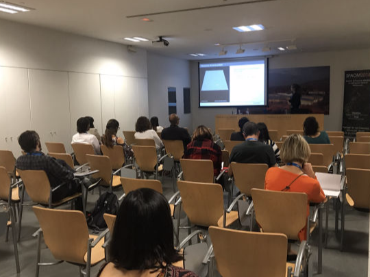
Several sessions which address key topics from image processing to advanced imaging, prepared and presented by your peers

Industry

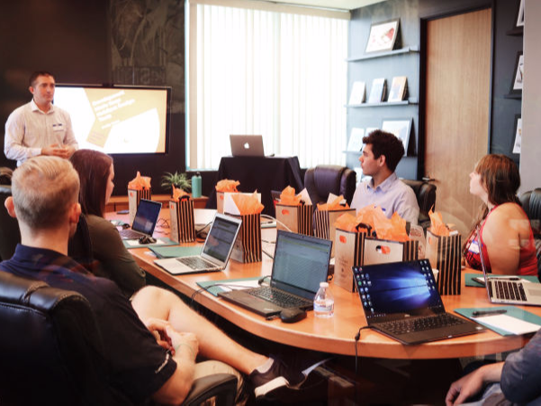
Series of presentations on technical advances and applications directly from manufacturers, will occur in a dedicated room

Community Workshops

Speakers:


Pedro Matos Pereira, ITQB NOVA
Carolina Feliciano, ITQB NOVA


Session Description:

Expanding biology above diffraction

Despite the widespread use of Super-Resolution techniques, some of them easy and freely available (e.g. SRRF or eSRRF), most methods remain technically challenging and involve sophisticated hardware and/or software tools. In 2014, Expansion Microscopy was introduced as an alternative to overcome this challenge. The concept is extremely simple, instead of super-resolving your target of interest you physically expand your sample in such a way that your structure is now above the diffraction limit (up to 10-fold the original size) and image it on a conventional microscope. The original method served to deliver super-resolution information to researchers without access to a super-resolution system. However, ExM’s versatility was limited by technical details like the need for special labelling probes. Several improvements were made since then to enhance its applicability. In this workshop we will show you how you can expand your super-resolution toolkit and your biology with it using expansion microscopy!

Speakers:


Davide Accardi, ABBE Platform, CCU

Pedro Campinho, ABBE Platform, CCU


Session Description:

The majority of biological processes happen in a three-dimensional space and some of them occur at a speed that would require multiple observations per second in order to be correctly followed. Because of their low phototoxicity, light-sheet systems have been recognized among the best instruments to follow living processes (ref.: Method of the Year 2014. Nat Methods 12, 1 (2015). https://doi.org/10.1038/nmeth.3251). However, volumetric high frequency imaging is not supported by the majority of commercially available light-sheet based microscopes.

During this workshop we will show theoretically and practically demonstrate a method to overcome this limitation by using the ZEISS light-sheet 7 (also valid for the previous model LZ.1) that would not require any system modification. This method allows to image at cellular resolution keeping the intrinsic confocality given by the system, yet imaging volumes between 50 and  100μm up to 33 times per second.


European Marine Biological Resource Centre (EMBRC): www.embrc.eu
EMBRC is Europe’s ‘research infrastructure’ for marine biological resources. This workshop is co-organised by the Portuguese and French nodes of EMBRC; EMBRC-PT and EMBRC-FR.


1. Shedding light on marine organisms

Speakers:
Sandra Silva, Rute Félix and Teresa Correia
CCMAR Algarve & EMBRC PT


Session Description:


Marine organisms encompass an outstanding range of biodiversity with valuable resources in both fundamental and applied research. The physiology of some marine species is greatly unknown and there are fewer molecular tools available. While several of them produce abundant eggs, embryos, and larvae that possess natural transparency making them readily available for microscopy, this is not the case of the majority of species with ecological or evolutionary interest. Current sample-clearing techniques are optimized for model organisms leaving out these marine species. In this workshop, we will dive into the marine organisms' research potential and the efforts that have been made to improve tissue clearing methods and develop molecular tools.

2. Marine microscopy developments: super-resolution and hyperspectral imaging

Speakers:

Sebastien Schaub, Institut de la Mer de Villefranche, Sorbonne Université & EMBRC France


Session Description:


Microscopy tools for marine sample imaging are sometimes similar to any microscopy platform and sometimes induces specificities.
In the first part, we will present a generic development about super-resolution. If some super-resolution techniques require either complex sample preparation or highly specific (and expensive) instruments, the methods based on fluorescence fluctuations are less demanding with a relative resolution improvement. Here we will present some result of COLORME and FLUOGAN we have developed and some illustrations on Ostreopsis phytoplankton which let us use standard microscopes (widefield or confocal).

In a second part, we will present Hyperspectral imaging results. Using a spectral detector with a white laser, we get 2D spectral imaging. A major difficulty in marine sample imaging is the autofluorescence which can easily crosstalk with exogenous stainings. For this reason, we develop this method to characterize the autofluorescence. We present some preliminary data on several plankton species and how it would let a better understanding and improve our sample staining. 
 

Speakers:


Jesús A. Andrés San Román, Departamento de Biología Celular, Universidad de Sevilla, Instituto de Biomedicina de Sevilla (IBiS)


Session Description:

A major bottleneck in the development of neural networks for cell segmentation is the requirement for labor-intensive manual curation to create a training dataset. This issue will be addressed in this workshop, along with the solution proposed by the Complex Organization of Living Matter Lab (@scu_lab): CartoCell.
CartoCell is a five-step pipeline for segmenting epithelial tissues that enables the quantification of geometric and packing features at the cellular level. The significant advantage of CartoCell is the ability to train with a minimal number of samples, thereby reducing the requirement for manual labor. Furthermore this pipeline can be easily utilized even without coding experience via Google Colab.
During this workshop, we will not only discuss the issue at hand but also provide a demonstration of how to use CartoCell to segment various types of epithelial tissues. 

Speakers:


Marcos Obando, Instituto Balseiro, Bariloche, Argentina
Giorgia Tortora, Politecnico di Milano, Italy


Session Description:

In the rapidly evolving fields of imaging and microscopy, identifying and developing accurate and efficient tools to analyse image data has become fundamental. In this context, napari emerges as a promising solution, providing a versatile platform for enhancing and expediting the image analysis process. napari is an interactive viewer designed for multi-dimensional images in Python which allows to visualise, annotate, and analyse images in a very easy and user-friendly way thanks to its layered structure and to all the image analysis plugins that it offers.
This workshop aims to acquaint participants with the fundamental aspects of napari. We will begin by familiarizing participants with the essential components of the napari viewer showing how to navigate and utilize napari interface efficiently. We will then show how to effectively exploit napari plugins by showing some cases that may be of interest for participants. Finally, considering that napari’s strength lies in the possibility to extend its capabilities through the creation of custom widgets, in the final part of the workshop we will explore how to create custom widgets within napari, tailored to users’ specific imaging needs.
This workshop not only serves as an introduction to napari but also as an invitation to join the community of researchers and scientists who contributes every day to expand the potential of napari in the world of imaging and microscopy. 
Speakers:


Johanna Bischof,  Euro-BioImaging


Session Description:

Euro-BioImaging is the European Infrastructure for Biological and Biomedical Imaging. With more than 170 imaging facilities across Europe, Euro-BioImaging provides open access to the full range of different imaging technologies across all scales as well as image data services. In this workshop we offer an opportunity to get to know Euro-BioImaging and our services, including presentations from some Nodes and facilities. We will also present different funding opportunities to support access to Euro-BioImaging services and other ways to get involved and benefit from Euro-BioImaging. 

Industry Workshops 

High Sensitivity Detection in Super Resolution: new NSPARC detector


Izasa will share two different locations where the lates technology in Advance Optical Microscopy will be presented.
In our booth the Nikon AX-confocal from with the latest technology in Super Resolution (NSPARC) and the biggest FOV of the market is waiting for you.

Also in the booth the unique spinning disk solution form Andor, the BC43 is performing the best images at the push of a button. Compact, fast and easy to use.

In the building 2 you will be surprised by the uniformity of the multicolor TIRF and HILO images that can be obtained at high speed with the Ilas2.

Improve your imaging of thick samples by using slide scanning with structured illumination


Widefield (WF) fluorescence microscopes provide high-resolution imaging for thin samples but are limited in their ability to image thicker samples due to background fluorescence causing image blur. Optical sectioning effectively overcomes this problem but has traditionally required a confocal system or deblurring software. With optical sectioning technology now on slide scanners, researchers can benefit from confocal capabilities for imaging thick samples—with significantly improved throughput.
High-Contrast Optical Sectioning Images :

The SILA (Speckle Illumination Acquisition) optical sectioning device easily integrates into your VS200 slides scanning system and software. Speckles are used to obtain high-contrast images, removing out-of-focus light to deliver sharp images, especially from thick samples. The image is computed during the scan, and there’s no need for post-processing, so the device has a minimal impact on acquisition speed. The SILA device—developed by Bliq Photonics—is a camera-based optical sectioning technology used to bring high-contrast, blur-free images to your VS200 system.

Easy to Use, The SILA device is easy to use with samples of any thickness and independent of the magnification. No special calibration is required, and it’s compatible with a variety of existing Evident objectives. Just insert the slide manually or using the loader, load the software, and acquire the images with the ‘SILA’ option enabled. Bring your slides and discover the significant benefits of the SILA optical sectioning device !!!


REGISTER HERE : https://www.zeiss.es/microscopia/l/eventos/registro-zeiss-a-spaom2023.html

25/10/2023

IWS3.1 » 14:45-15:30

Gauss and Bessel: Brothers in Arms for Exciting Samples in Lightsheet Microscopy, Jacques Paysan, ZEISS

IWS3.2  » 15:30-16:15

3D X-ray Microscopy (XRM) of Life Science specimens: visualize internal structures from microns to millimetres, Abel Sainz-Serrano, ZEISS

26/10/2023

IWS3.3 » 14:45-15:30

Your Complete Solution from Sample to Knowledge,
Verona Villar, ZEISS

IWS3.4 » 15:30-16:15

Hands on workshop: how to use AI in your image analysis pipelines, Delisa Garcia, ZEISS **

** Please bring your own Labtop – minimum requirements: Win10Pro 64bit/4-corex 86-compatilble CPU/8Gb RAM (16 GB recommended) GPU with support for OpenGL 4.3 or above and 2GB of VRAM (eg. Nvidia GeForce 1050 /ATI Radeon RX560) / 100GB spare local Storage
 

You can register here for the workshops:
    
      https://calendly.com/leica_microsystems/leica-workshops-at-spaom-2023 


25/10/2023

IWS4.1 » 14:45-15:30

Move into the next dimension with Stellaris and TauSense fluorescence lifetime imaging

IWS4.2 » 15:30-16:15

Autonomous imaging of rare events powered by Aivia AI and Stellaris

26/10/2023

IWS4.3 » 14:45-15:30

Move into the next dimension with Stellaris and TauSense fluorescence lifetime imaging

IWS4.4 » 15:30-16:15

Autonomous imaging of rare events powered by Aivia AI and Stellaris


© Copyright 2023 SPAOM - All Rights Reserved